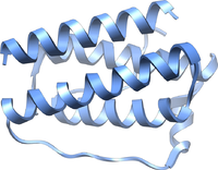
Photo from wikipedia
Chondrocytes in injured cartilage tissue are susceptible to mechanical loading; mechanical overloading can induce cartilage degeneration. The aim of the present study was to investigate whether mechanical loading can regulate… Click to show full abstract
Chondrocytes in injured cartilage tissue are susceptible to mechanical loading; mechanical overloading can induce cartilage degeneration. The aim of the present study was to investigate whether mechanical loading can regulate chondrocyte degeneration and angiogenesis via the tissue inhibitor of matrix metalloproteinase-3 (TIMP3)/transforming growth factor (TGF)-β1 axis. Primary human chondrocytes were obtained from knee articular cartilage of a healthy donor. Then, normal chondrocytes or TIMP3 lentivirus-transfected (LV-TIMP3) chondrocytes were subjected to mechanical loading (10 MPa compression). Then, chondrocytes were stimulated with 1 µg/ml lipopolysaccharide (LPS) or treated with LDN-193189 (inhibitor of TGF-β1 signaling pathway). In addition, human umbilical vein endothelial cells (HUVECs) were co-cultured with chondrocytes or LV-TIMP3 chondrocytes. The expression levels of collagen-I, proteoglycan, TIMP3, TGF-β1, Smad2 and Smad3 were detected by reverse transcription-quantitative PCR and western blotting. Moreover, cell apoptosis and viability were determined using flow cytometry and MTT analysis, while cell migration was observed by Transwell assays. In addition, the vascular endothelial growth factor (VEGF)/VEGF receptor (R)2 binding rate in HUVECs was detected by a solid-phase binding assay. It was demonstrated that mechanical loading significantly inhibited the expression levels of collagen-I and proteoglycan in chondrocytes, as well as reducing cell proliferation and promoting cell apoptosis. In addition, the expression levels of TIMP3, TGF-β1, phosphorylated (p)-Smad2 and p-Smad3 were significantly decreased in degenerated chondrocytes that were induced by LPS, as well as in chondrocytes treated with LDN-193189. Furthermore, TIMP3 overexpression suppressed cell migration and reduced the VEGF/VEGFR2 binding rate in HUVECs. Mechanical loading significantly inhibited the expression levels of TIMP3, TGF-β1, p-Smad2 and p-Smad3 in chondrocytes, and also increased cell migration of HUVECs; TGF-β1 treatment or TIMP3 overexpression reversed these effects. Thus, the TIMP3/TGF-β1 axis may be a vital signaling pathway in mechanical loading-induced chondrocyte degeneration and angiogenesis.
Journal Title: Molecular Medicine Reports
Year Published: 2020
Link to full text (if available)
Share on Social Media: Sign Up to like & get
recommendations!