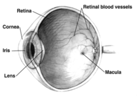
Photo from wikipedia
PURPOSE To report optical coherence tomography (OCT) of cherry-red spots from Tay-Sachs and Niemann-Pick disease. METHODS Consecutive patients with Tay-Sachs and Niemann-Pick disease evaluated by the pediatric transplant and cellular… Click to show full abstract
PURPOSE To report optical coherence tomography (OCT) of cherry-red spots from Tay-Sachs and Niemann-Pick disease. METHODS Consecutive patients with Tay-Sachs and Niemann-Pick disease evaluated by the pediatric transplant and cellular therapy team, for whom a handheld OCT scan was obtained, were included. Demographic information, clinical history, fundus photography, and OCT scans were reviewed. Two masked graders evaluated each of the scans. RESULTS The study included 3 patients with Tay-Sachs disease (5, 8, and 14 months old) and 1 patient with Niemann-Pick disease (12 months old). All patients had bilateral cherry-red spots on fundus examination. In all patients with Tay-Sachs disease, handheld OCT revealed parafoveal ganglion cell layer (GCL) thickening, increased nerve fiber layer, and GCL reflectivity, and different levels of residual normal signal GCL. The patient with Niemann-Pick disease had similar parafoveal findings, but there was a thicker residual GCL. Sedated visual evoked potentials were unrecordable in all 4 patients despite 3 of them demonstrating normal visual behavior for age. Patients with good vision had relative sparing of the GCL on OCT. CONCLUSIONS The cherry-red spots in lysosomal storage diseases appear as perifoveal thickening and hyperreflectivity of the GCL on OCT. In this case series, residual GCL with normal signal proved to be a better biomarker for visual function than visual evoked potentials and could be considered for future therapeutic trials. [J Pediatr Ophthalmol Strabismus. 20XX;X(X):XX-XX.].
Journal Title: Journal of pediatric ophthalmology and strabismus
Year Published: 2023
Link to full text (if available)
Share on Social Media: Sign Up to like & get
recommendations!