Photo from wikipedia
Alzheimer’s disease (AD) is a neurodegenerative disorder characterized by progressive cognitive impairment suggested to be induced by the accumulation of amyloid-β (Aβ) in the brain, especially in the hippocampus. Cerebral… Click to show full abstract
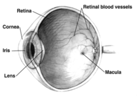
Alzheimer’s disease (AD) is a neurodegenerative disorder characterized by progressive cognitive impairment suggested to be induced by the accumulation of amyloid-β (Aβ) in the brain, especially in the hippocampus. Cerebral Aβ deposits may be detected through positron emission tomography (PET) as early as two decades before cl inically diagnosed ADassociated dementia, which provides the opportunity for early therapeutic interventions (Wang and Mao, 2021). PET may not be suitable for AD screening since it is invasive, costly, and inaccessible for routine clinical use or population screening. Aβ deposits have also been identified throughout the retina, which is a developmental outgrowth of the diencephalon and shares physiological and pathological pathways with the central nervous system (London et al., 2013). Patients with mild cognitive impairment and early AD are reported to have visual disturbances involving visual field loss with reported thinning of the retinal layers including the retinal nerve fiber layer, ganglion cell layer, and inner plexiform layer (KoronyoHamaoui et al., 2011; Wang and Mao, 2021). Retinal Aβ deposits have been detected prior to the manifestation of cerebral Aβ deposits in transgenic mice models of AD (KoronyoHamaoui et al., 2011; Habiba et al., 2021). Since the retina provides an easily accessible location for non-invasive imaging, retinal Aβ may have the potential to be a surrogate for cerebral Aβ and a biomarker for the detection of AD prior to irreversible cognitive impairment. Several techniques have been explored for imaging retinal Aβ, including the use of curcumin and hyperspectral imaging, which have been shown to differentiate AD patients and normal subjects in vivo (Koronyo et al., 2017; Hadoux et al., 2019). These non-invasive, imaging studies have also characterized retinal Aβ in human subjects and found correlations between retinal Aβ and cerebral manifestations including increased cerebral Aβ load and low cognitive assessment scores (Hadoux et al., 2019; Dumitrascu et al., 2020). However, further investigations with larger sample sizes and longitudinal studies are needed to determine if retinal Aβ can be applied in routine clinical settings and potentially for population-based screening.
Journal Title: Neural Regeneration Research
Year Published: 2022
Link to full text (if available)
Share on Social Media: Sign Up to like & get
recommendations!