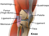
Photo from wikipedia
Introduction Trochlear dysplasia (TD) is a condition that is characterized by the presence of either a flat or convex trochlear, which impedes the stability of the patellofemoral joint (PFJ). The… Click to show full abstract
Introduction Trochlear dysplasia (TD) is a condition that is characterized by the presence of either a flat or convex trochlear, which impedes the stability of the patellofemoral joint (PFJ). The PFJ function is dependent on many different structures that surround the knee joint. The aim of this study was to analyse all the muscle components around the PFJ and identify whether gross muscle imbalance could contribute to the stability of the patella in TD. Material and methods The average cross-sectional area (CSA) and cross-sectional area ratio (CSAR) of each muscle of the thigh region in subtypes of TD was evaluated and compared to normal knee joints. Ninety-eight patients (196 knees in total) were included in the study. Results Of the 196 knee joints that were reviewed, 10 cases were found to be normal. In total, 186 cases were positive for TD. The majority consisted of type C. The hamstring muscles showed variable results. The vastus medialis muscle was larger in comparison to the vastus lateralis muscle over all the different TD subtypes; however, no statistical significance was identified. There was a marked statistical significance between the quadriceps and hamstring muscles, especially when comparing this to the normal knees within our cohort. Conclusions This study revealed no significant difference in the effect of the thigh muscle CSA on the stability of the PFJ in TD. Further research is required to establish the roles of the different muscles around PFJ in the prevention of TD dislocation.
Journal Title: Polish Journal of Radiology
Year Published: 2021
Link to full text (if available)
Share on Social Media: Sign Up to like & get
recommendations!