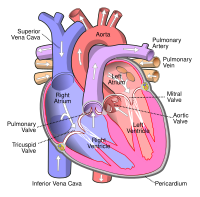
Photo from wikipedia
A 68-year-old man was admitted to our institution with dyspnea on exertion. Chest radiograph showed marked cardiomegaly. Transthoracic echocardiography displayed 2 parallel interatrial septa forming a distinct interatrial chamber, measuring… Click to show full abstract
A 68-year-old man was admitted to our institution with dyspnea on exertion. Chest radiograph showed marked cardiomegaly. Transthoracic echocardiography displayed 2 parallel interatrial septa forming a distinct interatrial chamber, measuring 2.4 cm × 0.8 cm (Figure 1A, Video 1). Color Doppler flow imaging showed a systolic flow into the interatrial chamber (Figure 1B) and severe mitral regurgitation owing to posterior mitral leaflet prolapse was noted (Figure 1C). Contrast echocardiography revealed the microbubbles entering into the interatrial chamber (Figure 1D, Video 2). Computed tomography angiography revealed the presence of a double atrial septum (DAS) with an interatrial accessory chamber (Figure 1E). For a better assessment of the interatrial septum, transesophageal echocardiography (TEE) was performed. Two-dimensional and 3-dimensional TEE demonstrated a double-layer membrane structure of the atrial septum and the distinct interatrial chamber communicating with the left atrium (Figures 1F-2B, Videos 3, 4). TrueVue images clearly exhibited the subtle structure of DAS with persistent interatrial space, making the images more closely resemble the real
Journal Title: Anatolian Journal of Cardiology
Year Published: 2022
Link to full text (if available)
Share on Social Media: Sign Up to like & get
recommendations!