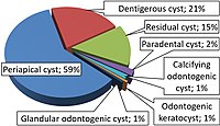
Photo from wikipedia
An 8 yr old female spayed golden retriever presented for a 3 wk history of progressive pelvic limb ataxia. MRI revealed a well-circumscribed T2-weighted hyperintense, T1-weighted poorly contrast-enhancing extradural mass… Click to show full abstract
An 8 yr old female spayed golden retriever presented for a 3 wk history of progressive pelvic limb ataxia. MRI revealed a well-circumscribed T2-weighted hyperintense, T1-weighted poorly contrast-enhancing extradural mass to the right of the spinal cord at the level of L1 causing severe spinal cord compression. A right-sided hemilaminectomy was performed to remove the mass, and histopathology revealed an intraosseous keratinized cyst. A complete neurologic recovery was made within 2 wk following the surgery. This case illustrates a rare diagnosis and the first case report describing MRI findings and favorable clinical outcome after surgical management of a spinal intraosseous keratinized cyst.
Journal Title: Journal of the American Animal Hospital Association
Year Published: 2022
Link to full text (if available)
Share on Social Media: Sign Up to like & get
recommendations!