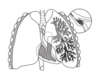
Photo from wikipedia
Various imaging modalities include chest computed tomography pulmonary angiography (CTPA), echocardiography, magnetic resonance imaging, and nuclear imaging and each are used for the assessment of varying status of PE. Assessment… Click to show full abstract
Various imaging modalities include chest computed tomography pulmonary angiography (CTPA), echocardiography, magnetic resonance imaging, and nuclear imaging and each are used for the assessment of varying status of PE. Assessment of thromboembolic burden by chest CTPA is the first step in the diagnosis of PE. Hemodynamic assessment can be achieved by echocardiography and also by chest CTPA. Nuclear imaging is useful in discriminating CTEPH from APE. Better perspectives on diagnosis, risk stratification and decision making in PE can be provided by combining multimodality cardiovascular imaging. Here, the advantages or pitfalls of each imaging modality in diagnosis, risk stratification, or management of PE will be discussed.
Journal Title: Cardiology journal
Year Published: 2019
Link to full text (if available)
Share on Social Media: Sign Up to like & get
recommendations!