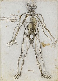
Photo from wikipedia
BACKGROUND We present a case report of double-headed extensor hallucis longus (EHL) with potential clinical significance. MATERIALS AND METHODS Cadaveric dissection of the right lower limb of a 70-year-old female… Click to show full abstract
BACKGROUND We present a case report of double-headed extensor hallucis longus (EHL) with potential clinical significance. MATERIALS AND METHODS Cadaveric dissection of the right lower limb of a 70-year-old female at death was performed for research and teaching purposes at the Department of Anatomical Dissection and Donation, Medical University of Lodz. The limb was dissected using standard techniques according to a strictly specified protocol. Each head and tendon of the muscle was photographed and subjected to further measurements. RESULTS During dissection, an unusual type of EHL muscle was observed. It consisted of two muscle bellies, a main tendon and an accessory tendon. Both muscle bellies were located on anterior surface of the fibula and the interosseous membrane. The main tendon insertion was located on the dorsal aspect of the base of the distal phalanx of the big toe, while the accessory tendon insertion was located medially. CONCLUSIONS The EHL muscle is highly morphologically variable at both the point of origin and the insertion. Knowledge of its variationsis connected to several pathologies such as foot drop, tendonitis, tendon rupture, and anterior compartment syndrome.
Journal Title: Folia morphologica
Year Published: 2022
Link to full text (if available)
Share on Social Media: Sign Up to like & get
recommendations!