Photo from wikipedia
AIM OF THE STUDY Tumours of the infratemporal fossa (ITF) are rare and include primary tumours, contiguity lesions and metastases. Surgical resection is the gold standard. The fronto-orbito-zygomatic (FOZ) approach… Click to show full abstract
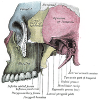
AIM OF THE STUDY Tumours of the infratemporal fossa (ITF) are rare and include primary tumours, contiguity lesions and metastases. Surgical resection is the gold standard. The fronto-orbito-zygomatic (FOZ) approach is commonly used in order to obtain safe access to the lateral skull base and ITF to resect intra- and extra-cranial tumours. We here describe our series of ITF lesions extending to the middle cranial fossa and/or orbit, treated by single- or two piece FOZ. MATERIAL AND METHODS All cases of single- or two-piece FOZ approach for an infratemporal fossa lesion extending to the middle cranial fossa operated at our Institution from January 2014 to January 2018 were retrospectively reviewed. The follow-up was for a minimum of four months and a maximum of 60 months. The inclusion criteria were lesions involving the ITF with an extension to the middle cranial fossa and/or orbit. Baseline characteristics of patients, tumour localisation, tumour extension, diffusion route, histology, extent of tumour resection, postoperative treatment, and post-operative complications were evaluated. RESULTS Nine patients underwent a surgical procedure with a FOZ approach, two of them with a single-piece approach and the remainder with a two-piece one. All patients had an ITF localisation. Gross total removal (GTR) was achieved in 7/9 patients. Only one patient, with non-total removal (NTR), underwent radiotherapy. CONCLUSIONS For the treatment of ITF fossa tumours extending to the orbit and or middle cranial fossa, we believe that both FOZ techniques are effective and allow a good medial extension toward the cavernous sinus and parasellar region. But a two-piece craniotomy may ensure a more medial extension and a wider angle of work compared to a one-piece craniotomy.
Journal Title: Neurologia i neurochirurgia polska
Year Published: 2022
Link to full text (if available)
Share on Social Media: Sign Up to like & get
recommendations!