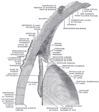
Photo from wikipedia
PURPOSE We aimed to evaluate the use of automated quantitative static and dynamic pupillometry in screening patients with type 2 diabetes mellitus and different stages of diabetic retinopathy. METHOD 155… Click to show full abstract
PURPOSE We aimed to evaluate the use of automated quantitative static and dynamic pupillometry in screening patients with type 2 diabetes mellitus and different stages of diabetic retinopathy. METHOD 155 patients with type 2 diabetes mellitus (diabetes mellitus group) were included in this study and another 145 age- and sex-matched healthy individuals to serve as the control group. The diabetes mellitus group was divided into three subgroups: diabetes mellitus without diabetic retinopathy (No-diabetic retinopathy), nonproliferative diabetic retinopathy, and proliferative diabetic retinopathy. Static and dynamic pupillometry were performed using a rotating Scheimpflug camera with a topography-based system. RESULTS In terms of pupil diameter in both static and dynamic pupillometry (p<0.05), statistically significant differences were observed between the diabetes mellitus and control groups and also between the subgroups No-diabetic retinopathy, nonproliferative diabetic retinopathy, and proliferative diabetic retinopathy subgroups. But it was noted that No-diabetic retinopathy and nonproliferative diabetic retinopathy groups have showed similarities in the findings derived from static pupillometry under mesopic and photopic conditions. The two groups also appeared similar at all points during the dynamic pupillometry (p>0.05). However, it could be concluded that the proliferative diabetic retinopathy group was significantly different from the rest of the subgroups, No-diabetic retinopathy and nonproliferative diabetic retinopathy groups, in terms of all the static pupillometry measurements (p<0.05). The average speed of dilation was also significantly different between the diabetes mellitus and control groups and among the diabetes mellitus subgroups (p<0.001). While weak to moderate significant correlations were found between all pupil diameters in static and dynamic pupillometry with the duration of diabetes mellitus (p<0.05 for all), the HbA1c values showed no statistically significant correlations with any of the investigated static and dynamic pupil diameters (p>0.05 for all). CONCLUSION This study revealed that the measurements derived from automated pupillometry are altered in patients with type 2 diabetes mellitus. The presence of nonproliferative diabetic retinopathy does not have a negative effect on pupillometry findings, but with proliferative diabetic retinopathy, significant alterations were observed. These results suggest that using automated quantitative pupillometry may be useful in verifying the severity of diabetic retinopathy.
Journal Title: Arquivos brasileiros de oftalmologia
Year Published: 2021
Link to full text (if available)
Share on Social Media: Sign Up to like & get
recommendations!