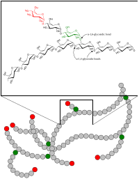
Photo from wikipedia
To date, microglia subsets in the healthy CNS have not been identified. Utilizing autofluorescence (AF) as a discriminating parameter, we identified two novel microglia subsets in both mice and non-human… Click to show full abstract
To date, microglia subsets in the healthy CNS have not been identified. Utilizing autofluorescence (AF) as a discriminating parameter, we identified two novel microglia subsets in both mice and non-human primates, termed autofluorescence-positive (AF+) and negative (AF−). While their proportion remained constant throughout most adult life, the AF signal linearly and specifically increased in AF+ microglia with age and correlated with a commensurate increase in size and complexity of lysosomal storage bodies, as detected by transmission electron microscopy and LAMP1 levels. Post-depletion repopulation kinetics revealed AF− cells as likely precursors of AF+ microglia. At the molecular level, the proteome of AF+ microglia showed overrepresentation of endolysosomal, autophagic, catabolic, and mTOR-related proteins. Mimicking the effect of advanced aging, genetic disruption of lysosomal function accelerated the accumulation of storage bodies in AF+ cells and led to impaired microglia physiology and cell death, suggestive of a mechanistic convergence between aging and lysosomal storage disorders.
Journal Title: eLife
Year Published: 2020
Link to full text (if available)
Share on Social Media: Sign Up to like & get
recommendations!