Transcranial color‐coded sonography may indirectly reveal an intracranial meningioma
Sign Up to like & getrecommendations! Published in 2019 at "Journal of Clinical Ultrasound"
DOI: 10.1002/jcu.22737
Abstract: Meningiomas are extra‐axial tumors with a long‐standing indolent clinical course. Sphenoid wing meningiomas may slowly grow, spreading toward the orbitofrontal and temporal regions as well as encasing the middle cerebral artery (MCA). Herein, we report… read more here.
Keywords: coded sonography; sonography may; meningioma; transcranial color ... See more keywords

Color-coded Digital Subtraction Angiography for Assessing Acute Skeletal Muscle Ischemia-Reperfusion Injury in a Rabbit Model.
Sign Up to like & getrecommendations! Published in 2018 at "Academic radiology"
DOI: 10.1016/j.acra.2018.03.020
Abstract: RATIONALE AND OBJECTIVES This paper describes an ongoing investigation of imaging and characterization of ischemia-reperfusion (IR) and investigated the use of color-coded digital subtraction angiography (DSA) to assess reperfusion injury or potential injury. METHODS New… read more here.
Keywords: muscle; peak; reperfusion; color coded ... See more keywords

Transcranial color-coded sonography of vertebral artery for diagnosis of right-to-left shunts
Sign Up to like & getrecommendations! Published in 2017 at "Journal of the Neurological Sciences"
DOI: 10.1016/j.jns.2017.03.012
Abstract: BACKGROUND It is unknown whether contrast transcranial color-coded sonography of vertebral artery monitoring via the foramen magnum window (cTCCS-VA) is useful to detect right-to-left shunt (RLS). We investigated whether cTCCS-VA can be proposed as an… read more here.
Keywords: coded sonography; artery; diagnosis; color coded ... See more keywords

In reply to "Bias in the interpretation of the transcranial color-coded duplex sonography register".
Sign Up to like & getrecommendations! Published in 2019 at "Medicina intensiva"
DOI: 10.1016/j.medin.2018.08.006
Abstract: We wish to thank the interest shown by Rochetti and Egea-Guerrero toward our paper ‘‘Desaparición del flujo diastólico cerebral tras una complicación inesperada’’ (Disappearance of diastolic flow after unexpected complication) and their interesting comments made… read more here.
Keywords: coded duplex; duplex sonography; color coded; flow ... See more keywords

Color-coded intravital imaging demonstrates a transforming growth factor-β (TGF-β) antagonist selectively targets stromal cells in a human pancreatic-cancer orthotopic mouse model
Sign Up to like & getrecommendations! Published in 2017 at "Cell Cycle"
DOI: 10.1080/15384101.2017.1315489
Abstract: ABSTRACT Pancreatic cancer is a recalcitrant malignancy, partly due to desmoplastic stroma which stimulates tumor growth, invasion, and metastasis, and inhibits chemotherapeutic drug delivery. Transforming growth factor-β (TGF-β) has an important role in the formation… read more here.
Keywords: pancreatic cancer; model; color coded; cancer ... See more keywords

Color-coded computer-generated Moiré profilometry with real-time 3D measurement and synchronous monitoring video collection
Sign Up to like & getrecommendations! Published in 2021 at "Optical Engineering"
DOI: 10.1117/1.oe.60.3.034109
Abstract: Abstract. A color-coded computer-generated Moiré profilometry (CGMP) with real-time 3D measurement and synchronous monitoring video collection is proposed. In this method, a sinusoidal grating and the direct current (DC) component of it are encoded in… read more here.
Keywords: deformed pattern; real time; monitoring; color ... See more keywords

Reliability and Accuracy of Peri-Interventional Stenosis Grading in Peripheral Artery Disease Using Color-Coded Quantitative Fluoroscopy: A Phantom Study Comparing a Clinical and Scientific Postprocessing Software
Sign Up to like & getrecommendations! Published in 2018 at "BioMed Research International"
DOI: 10.1155/2018/6180138
Abstract: Purpose To assess quantitative stenosis grading by color-coded fluoroscopy using an in vitro pulsatile flow phantom. Methods Three different stenotic tubes (80%, 60%, and 40% diameter restriction) and a nonstenotic reference tube were compared regarding… read more here.
Keywords: stenosis grading; grade; stenosis; color coded ... See more keywords

Color-coded summation images in the evaluation of renal artery stenosis before and after percutaneous transluminal angioplasty
Sign Up to like & getrecommendations! Published in 2021 at "BMC Medical Imaging"
DOI: 10.1186/s12880-020-00540-w
Abstract: Background Endovascular therapy is the gold standard in patients with hemodynamic relevant renal artery stenosis (RAS) resistant to medical therapy. The severity grading of the stenosis as well as the result assessment after endovascular approach… read more here.
Keywords: artery stenosis; renal artery; artery; color coded ... See more keywords

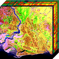
Adaptive filter design via a gradient thresholding algorithm for compressive spectral imaging.
Sign Up to like & getrecommendations! Published in 2018 at "Applied optics"
DOI: 10.1364/ao.57.004890
Abstract: Sensing a spectral image data cube has traditionally been a time-consuming task since it requires a scanning process. In contrast, compressive spectral imaging (CSI) has attracted widespread interest since it requires fewer samples than scanning… read more here.
Keywords: data cube; compressive spectral; spectral imaging; color coded ... See more keywords

Visualizing the Tumor Microenvironment by Color-coded Imaging in Orthotopic Mouse Models of Cancer.
Sign Up to like & getrecommendations! Published in 2018 at "Anticancer research"
DOI: 10.21873/anticanres.12423
Abstract: The tumor microenvironment (TME) contains stromal cells in a complex interaction with cancer cells. This relationship has become better understood with the use of fluorescent proteins for in vivo imaging, originally developed by our laboratories.… read more here.
Keywords: color; stromal cells; coded imaging; fluorescent protein ... See more keywords

Diagnostic performance of the thyroid imaging reporting and data system improved by color-coded acoustic radiation force pulse imaging.
Sign Up to like & getrecommendations! Published in 2023 at "Journal of X-ray science and technology"
DOI: 10.3233/xst-221359
Abstract: OBJECTIVE To explore the value of color-coded virtual touch tissue imaging (CCV) using acoustic radiation force pulse technology (ARFI) in diagnosing malignant thyroid nodules. METHODS Images including 189 thyroid nodules were collected as training samples… read more here.
Keywords: thyroid; color coded; rads ccv; diagnostic performance ... See more keywords