
Accelerating FLAIR imaging via deep learning reconstruction: potential for evaluating white matter hyperintensities
Sign Up to like & getrecommendations! Published in 2024 at "Japanese Journal of Radiology"
DOI: 10.1007/s11604-024-01666-5
Abstract: To evaluate deep learning-reconstructed (DLR)–fluid-attenuated inversion recovery (FLAIR) images generated from undersampled data, compare them with fully sampled and rapidly acquired FLAIR images, and assess their potential for white matter hyperintensity evaluation. We examined 30… read more here.
Keywords: white matter; dlr flair; flair; flair images ... See more keywords
Efficient segmentation of active and inactive plaques in FLAIR-images using DeepLabV3Plus SE with efficientnetb0 backbone in multiple sclerosis
Sign Up to like & getrecommendations! Published in 2024 at "Scientific Reports"
DOI: 10.1038/s41598-024-67130-6
Abstract: This research paper introduces an efficient approach for the segmentation of active and inactive plaques within Fluid-attenuated inversion recovery (FLAIR) images, employing a convolutional neural network (CNN) model known as DeepLabV3Plus SE with the EfficientNetB0… read more here.
Keywords: active inactive; segmentation; segmentation active; inactive plaques ... See more keywords
Slight hyperintensity of the left piriform cortex and amygdala on T2-weighted FLAIR images in older adults and patients with probable Alzheimer's disease
Sign Up to like & getrecommendations! Published in 2025 at "Acta Radiologica"
DOI: 10.1177/02841851251328261
Abstract: Background The left piriform cortex and amygdala (PC&A) is an early target for deterioration due to aging and Alzheimer's disease (AD) in several neuropathological and magnetic resonance (MR) volumetric studies. We observed slight hyperintensity of… read more here.
Keywords: cortex amygdala; piriform cortex; flair images; left piriform ... See more keywords
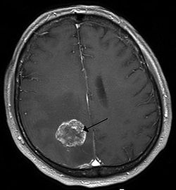
Preoperative FLAIR images for identifying glioblastoma boundaries
Sign Up to like & getrecommendations! Published in 2025 at "BMC Medical Imaging"
DOI: 10.1186/s12880-025-01839-2
Abstract: Glioblastoma is the most aggressive and rapidly growing type of central nervous system tumor. Despite advancements in imaging, no objective measurement for predicting the true extent of glioblastoma has been established. Compared with contrast-enhanced magnetic… read more here.
Keywords: glioblastoma; flair; preoperative flair; tumor ... See more keywords
Peritumoral Hyperintense Signal on Postcontrast FLAIR Images Surrounding Vestibular Schwannomas following Stereotactic Radiosurgery
Sign Up to like & getrecommendations! Published in 2025 at "American Journal of Neuroradiology"
DOI: 10.3174/ajnr.a8657
Abstract: BACKGROUND AND PURPOSE: Prior investigations have noted the presence of peritumoral hyperintense signal (a “halo”) around vestibular schwannomas on postcontrast 3D T2 FLAIR images. This study evaluated this phenomenon in a cohort of patients undergoing… read more here.
Keywords: halo; stereotactic radiosurgery; flair images; vestibular schwannomas ... See more keywords

Specialized gray matter segmentation via a generative adversarial network: application on brain white matter hyperintensities classification
Sign Up to like & getrecommendations! Published in 2024 at "Frontiers in Neuroscience"
DOI: 10.3389/fnins.2024.1416174
Abstract: Background White matter hyperintensities (WMH) observed in T2 fluid-attenuated inversion recovery (FLAIR) images have emerged as potential markers of neurodegenerative diseases like Multiple Sclerosis (MS). Lacking comprehensive automated WMH classification systems in current research, there… read more here.
Keywords: classification; white matter; method; wmh ... See more keywords
Hyperintensity of Cerebrospinal Fluid on T2-Weighted Fluid-Attenuated Inversion Recovery Magnetic Resonance Imaging Caused by High Inspired Oxygen Fraction
Sign Up to like & getrecommendations! Published in 2017 at "Frontiers in Veterinary Science"
DOI: 10.3389/fvets.2017.00219
Abstract: In veterinary medicine, patients undergo magnetic resonance imaging (MRI) under general anesthesia to enable acquisition of artifact-free images. The fraction of inspired oxygen (FiO2) ranges between 30 and 95%. In humans, a high FiO2 is… read more here.
Keywords: oxygen; fluid; flair images; t2w flair ... See more keywords