
Grayscale representation of infrared microscopy images by extended multiplicative signal correction for registration with histological images
Sign Up to like & getrecommendations! Published in 2020 at "Journal of Biophotonics"
DOI: 10.1002/jbio.201960223
Abstract: Fourier‐transform infrared (FTIR) microspectroscopy is rounding the corner to become a label‐free routine method for cancer diagnosis. In order to build infrared‐spectral based classifiers, infrared images need to be registered with Hematoxylin and Eosin (H&E)… read more here.
Keywords: multiplicative signal; microscopy; information; extended multiplicative ... See more keywords
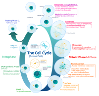
Efficient automated detection of mitotic cells from breast histological images using deep convolution neutral network with wavelet decomposed patches
Sign Up to like & getrecommendations! Published in 2019 at "Computers in biology and medicine"
DOI: 10.1016/j.compbiomed.2018.11.001
Abstract: In medical practice, the mitotic cell count from histological images acts as a proliferative marker for cancer diagnosis. Therefore, an accurate method for detecting mitotic cells in histological images is essential for cancer screening. Manual… read more here.
Keywords: mitotic cell; detection; automated detection; detection mitotic ... See more keywords

A new method for automatic counting of ovarian follicles on whole slide histological images based on convolutional neural network
Sign Up to like & getrecommendations! Published in 2019 at "Computers in biology and medicine"
DOI: 10.1016/j.compbiomed.2019.103350
Abstract: The ovary is a complex endocrine organ that shows significant structural and functional changes in the female reproductive system over recurrent cycles. There are different types of follicles in the ovarian tissue. The reproductive potential… read more here.
Keywords: images based; method; based convolutional; slide histological ... See more keywords

Robust extraction of quantitative structural information from high-variance histological images of livers from necropsied Soay sheep
Sign Up to like & getrecommendations! Published in 2017 at "Royal Society Open Science"
DOI: 10.1098/rsos.170111
Abstract: Quantitative information is essential to the empirical analysis of biological systems. In many such systems, spatial relations between anatomical structures is of interest, making imaging a valuable data acquisition tool. However, image data can be… read more here.
Keywords: structural information; information; high variance; image ... See more keywords

Pixel-Wise Classification in Hippocampus Histological Images
Sign Up to like & getrecommendations! Published in 2021 at "Computational and Mathematical Methods in Medicine"
DOI: 10.1155/2021/6663977
Abstract: This paper presents a method for pixel-wise classification applied for the first time on hippocampus histological images. The goal is achieved by representing pixels in a 14-D vector, composed of grey-level information and moment invariants.… read more here.
Keywords: pixel wise; histological images; wise classification; hippocampus histological ... See more keywords

Comparison in Quantities from Including Angles Comprising Lines of Hypha Themselves in Histological Images between Mucorales and Aspergillus.
Sign Up to like & getrecommendations! Published in 2019 at "Medical mycology journal"
DOI: 10.3314/mmj.19-00006
Abstract: BACKGROUND The rate of aspergillosis has decreased due to improvements in therapy. The rate of mucormycosis, however, has gradually increased in recent years. Both aspergillosis and mucormycosis produce histologically similar hyphae, pointing to the need… read more here.
Keywords: quantities including; intravascular lesions; aspergillosis; comparison quantities ... See more keywords