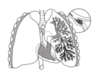
Quantitative lung perfusion blood volume (PBV) using dual energy CT (DECT)-based effective atomic number (Zeff ) imaging.
Sign Up to like & getrecommendations! Published in 2021 at "Medical physics"
DOI: 10.1002/mp.15227
Abstract: BACKGROUND Iodine material images (aka iodine basis images) generated from dual energy CT (DECT) have been used to assess potential perfusion defects in the pulmonary parenchyma. However, iodine material images do not provide the needed… read more here.
Keywords: perfusion defects; perfusion; iodine material; effective atomic ... See more keywords

Detection and quantitation of right ventricular reversible perfusion defects by stress SPECT myocardial perfusion imaging: A proof-of-principle study
Sign Up to like & getrecommendations! Published in 2017 at "Journal of Nuclear Cardiology"
DOI: 10.1007/s12350-017-0954-4
Abstract: BackgroundIn patients with right dominant coronary circulation, the right ventricular (RV) myocardium and the inferior region of the left ventricular (LV) myocardium share a common source of blood flow. We hypothesized that stress/rest SPECT myocardial… read more here.
Keywords: perfusion defects; perfusion; myocardial perfusion; reversible perfusion ... See more keywords

Right ventricular reversible perfusion defects
Sign Up to like & getrecommendations! Published in 2019 at "Journal of Nuclear Cardiology"
DOI: 10.1007/s12350-019-01697-w
Abstract: To the Editor, We thank Zhou et al. for their careful reading of our manuscript and for pointing out an error in the abstract. We agree that the age and gender were not statistically different… read more here.
Keywords: medicine; perfusion defects; cardiology; reversible perfusion ... See more keywords

Head-to-head comparison of lung perfusion with dual-energy CT and SPECT-CT.
Sign Up to like & getrecommendations! Published in 2020 at "Diagnostic and interventional imaging"
DOI: 10.1016/j.diii.2020.02.006
Abstract: PURPOSE To compare the quantitative and qualitative lung perfusion data acquired with dual energy CT (DECT) to that acquired with a large field-of-view cadmium-zinc-telluride camera single-photon emission CT coupled to a CT system (SPECT-CT). MATERIALS… read more here.
Keywords: head; perfusion defects; lung perfusion; perfusion ... See more keywords

Improved Discrimination of Myocardial Perfusion Defects at Low Energy Levels Using Virtual Monochromatic Imaging
Sign Up to like & getrecommendations! Published in 2017 at "Journal of Computer Assisted Tomography"
DOI: 10.1097/rct.0000000000000584
Abstract: Objectives The aim of this study was to explore the diagnostic performance of dual-energy computed tomography perfusion (DE-CTP) at different energy levels. Methods Patients with known or suspected coronary artery disease underwent stress and rest… read more here.
Keywords: perfusion defects; energy levels; perfusion; energy ... See more keywords

Mismatched perfusion defects on routine ventilation‐perfusion scans after lung transplantation
Sign Up to like & getrecommendations! Published in 2022 at "Clinical Transplantation"
DOI: 10.1111/ctr.14650
Abstract: Incidental pulmonary embolism (PE) is a challenging entity with unclear treatment implications. Our program performs routine ventilation‐perfusion (VQ) scans at 3‐months post‐transplant to establish airway and vascular function. We sought to determine the prevalence and… read more here.
Keywords: mismatched perfusion; perfusion defects; routine ventilation; perfusion scans ... See more keywords

Association between left ventricular perfusion defects and myocardial deformation indexes in heart transplantation recipients
Sign Up to like & getrecommendations! Published in 2017 at "Echocardiography"
DOI: 10.1111/echo.13596
Abstract: The aim of the study was to analyze possible correlations between strain echocardiography (STE) and PET myocardial perfusion in a population of heart transplantation (HTx) recipients showing preserved left ventricular (LV) ejection fraction. By STE,… read more here.
Keywords: perfusion defects; perfusion; htx; heart transplantation ... See more keywords

Apical Ischemia Is a Universal Feature of Apical Hypertrophic Cardiomyopathy
Sign Up to like & getrecommendations! Published in 2023 at "Circulation. Cardiovascular Imaging"
DOI: 10.1161/circimaging.122.014907
Abstract: Background: Apical hypertrophic cardiomyopathy (ApHCM) accounts for ≈10% of hypertrophic cardiomyopathy cases and is characterized by apical hypertrophy, apical cavity obliteration, and tall ECG R waves with ischemic-looking deep T-wave inversion. These may be present… read more here.
Keywords: hypertrophic cardiomyopathy; apical hypertrophic; hypertrophy; aphcm ... See more keywords

Prevalence and clinical significance of cardiovascular magnetic resonance adenosine stress-induced myocardial perfusion defect in hypertrophic cardiomyopathy
Sign Up to like & getrecommendations! Published in 2020 at "Journal of Cardiovascular Magnetic Resonance"
DOI: 10.1186/s12968-020-00623-1
Abstract: Background Hypertrophic cardiomyopathy (HCM) is thought to be associated with microvascular dysfunction. Adenosine stress-perfusion cardiovascular magnetic resonance imaging (CMR) is a sensitive method for assessing microvascular perfusion abnormalities. We evaluated the prevalence and clinical characteristics… read more here.
Keywords: perfusion defects; adenosine stress; perfusion; stress induced ... See more keywords

Comparison of dual-energy computer tomography and dynamic contrast-enhanced MRI for evaluating lung perfusion defects in chronic thromboembolic pulmonary hypertension
Sign Up to like & getrecommendations! Published in 2021 at "PLoS ONE"
DOI: 10.1371/journal.pone.0251740
Abstract: Objectives To evaluate the agreement in detecting pulmonary perfusion defects in patients with chronic thromboembolic pulmonary hypertension using dual-energy CT and dynamic contrast-enhanced MRI. Second, to compare both imaging modalities in monitoring lung perfusion changes… read more here.
Keywords: perfusion defects; chronic thromboembolic; lung perfusion; perfusion ... See more keywords

Lung Ventilation/Perfusion Scintigraphy for the Screening of Chronic Thromboembolic Pulmonary Hypertension (CTEPH): Which Criteria to Use?
Sign Up to like & getrecommendations! Published in 2022 at "Frontiers in Medicine"
DOI: 10.3389/fmed.2022.851935
Abstract: Objective The diagnosis of chronic thromboembolic pulmonary hypertension (CTEPH) is a major challenge as it is a curable cause of pulmonary hypertension (PH). Ventilation/Perfusion (V/Q) lung scintigraphy is the imaging modality of choice for the… read more here.
Keywords: pulmonary hypertension; scintigraphy screening; perfusion; perfusion defects ... See more keywords